Esophageal Varices: Causes, Symptoms, Diagnosis, Treatment, and Prevention
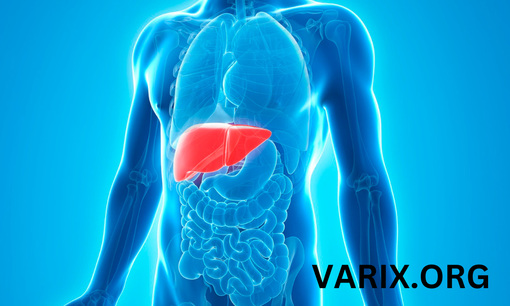
Esophageal varices are dilated veins in the lower part of the esophagus that develop in the setting of portal hypertension. Most often, they occur in people with severe liver disease such as cirrhosis, because scar tissue blocks normal blood flow through the liver. When pressure rises in the portal vein, blood is rerouted into smaller vessels that are not designed for high volume. This causes these blood vessels in the esophagus to enlarge and become fragile. Untreated esophageal varices can rupture and cause serious bleeding, which requires emergency treatment.
Causes and Risk Factors
The main cause of esophageal varices is longstanding liver disease that leads to portal hypertension. Risk factors include:
- Cirrhosis: Scarring of the liver from chronic hepatitis, alcoholic liver disease, or nonalcoholic fatty liver.
- Chronic hepatitis B or C: Viral infections that damage liver cells and lead to fibrosis.
- Alcoholic liver disease: Long-term alcohol abuse causes inflammation, fatty buildup, and scarring of the liver.
- Schistosomiasis: A parasitic infection that can affect the portal vein in endemic regions.
- Fatty liver disease: Nonalcoholic steatohepatitis (NASH) leads to cirrhosis in some patients.
Signs and Symptoms
Esophageal varices themselves often cause no symptoms until they bleed. When bleeding occurs, symptoms may include:
- Vomiting large amounts of bright red blood
- Black, tarry stools (melena)
- Lightheadedness or dizziness
- Low blood pressure and rapid heartbeat
- Loss of consciousness in severe cases
In the absence of bleeding, patients may have signs of underlying liver disease such as:
- Jaundice (yellowing of skin or eyes)
- Easy bruising or bleeding
- Abdominal swelling (ascites)
- Fatigue and weakness
Diagnosis of Esophageal Varices
Endoscopic Evaluation
The gold standard for detecting and grading esophageal varices is upper endoscopy. A flexible tube with a camera is inserted through the mouth to directly visualize the veins. Varices are graded based on size:
- Small: Thin, straight varices that do not protrude into the lumen.
- Medium: Tortuous veins occupying less than one-third of the esophageal lumen.
- Large: Bulging veins occupying more than one-third of the lumen with red wale marks indicating recent or imminent bleeding.
Imaging and Noninvasive Tests
In addition to endoscopy, the following studies can assess liver health and portal pressure:
- Ultrasound with Doppler: Evaluates blood flow in the portal vein and checks for ascites or splenomegaly.
- CT or MRI scan: Visualizes liver cirrhosis, nodules, and collateral vessels.
- Transient elastography (FibroScan): Measures liver stiffness to estimate fibrosis severity.
Treatment to Prevent Bleeding
Once esophageal varices are identified, the primary goal is to lower portal pressure and prevent variceal bleeding:
- Nonselective β-blockers: Medications such as propranolol or nadolol reduce portal flow by lowering heart rate and splanchnic circulation. They can decrease the risk of first bleed.
- Endoscopic variceal ligation (EVL): Elastic rubber bands are applied during endoscopy to ligate and obliterate varices. This is recommended for medium to large varices to prevent first or recurrent bleeding.
- Endoscopic sclerotherapy: Injection of a sclerosing agent directly into or adjacent to varices induces thrombosis and fibrosis. Typically used if ligation is not feasible.
- Transjugular intrahepatic portosystemic shunt (TIPS): A stent is placed between the portal vein and hepatic vein to decompress the portal system. Indicated for refractory or recurrent bleeding.
- Balloon tamponade: A temporary measure in emergency bleeding, using a Sengstaken-Blakemore tube to compress varices until definitive therapy is available.
Management of Acute Variceal Bleeding
Acute bleeding from esophageal varices is a life-threatening emergency. Key steps in management include:
- Resuscitation: Establish two large-bore IV lines, transfuse blood products, correct coagulopathy, and maintain airway protection.
- Vasoactive drugs: Initiate octreotide or terlipressin to constrict splanchnic vessels and reduce portal pressure.
- Emergency endoscopy: Perform as soon as the patient is hemodynamically stable. Use band ligation as first-line therapy for bleeding varices.
- Balloon tamponade or Sengstaken-Blakemore tube: Use only as a bridge to definitive therapy if endoscopic control is not possible.
- Antibiotic prophylaxis: Administer intravenous ceftriaxone or norfloxacin to reduce infection risk in cirrhotic patients.
- TIPS placement: Consider early in patients who fail endoscopic and pharmacologic therapy to control bleeding.
Prevention and Long-Term Management
Lifestyle and Dietary Measures
Although you cannot reverse established cirrhosis, certain actions can help reduce portal hypertension progression and lower variceal risk:
- Avoid alcohol completely if liver disease is alcohol-related.
- Maintain a healthy weight and balanced diet rich in fruits, vegetables, and lean proteins.
- Control viral hepatitis with antiviral therapy and regular monitoring.
- Engage in moderate exercise to improve circulation and overall health.
Regular Surveillance Endoscopy
Patients with cirrhosis should undergo screening endoscopy every 1–2 years to detect new or worsening varices. If small varices are found, repeat endoscopy is recommended in 1–2 years. For those with medium or large varices, start primary prophylaxis with β-blockers or EVL immediately.
Conclusion
Esophageal varices represent a serious complication of portal hypertension. Early detection through endoscopy, appropriate use of nonselective β-blockers and endoscopic band ligation, and lifestyle modifications can greatly reduce the risk of life-threatening bleeding. If bleeding occurs, prompt resuscitation, vasoactive therapy, and endoscopic intervention are critical. For patients with refractory variceal bleeding, TIPS placement provides an effective means of reducing portal pressure. Work closely with a hepatology or gastroenterology specialist to develop an individualized management plan.
Disclaimer: This article is intended for informational purposes only and does not constitute medical advice, diagnosis, or treatment. The content provided should not be used as a substitute for professional medical advice, diagnosis, or treatment. Always consult with a qualified healthcare professional before making any decisions about your health or medical conditions. Never disregard or delay seeking professional medical advice due to the information provided in this article. The author and publisher of this article are not responsible or liable for any adverse outcomes resulting from the use or reliance on the information provided herein.